Diagnosis of scleroderma tests. Treatment of systemic scleroderma.
Systemic scleroderma is a connective tissue pathology that affects the skin, musculoskeletal system, internal organs and blood vessels. The essence of the disease is that blood circulation is disturbed, connective tissue grows, becomes inflamed and thickened.
Systemic scleroderma refers to autoimmune diseases - this means malfunctions when it begins to attack the cells of its own body.
Scleroderma, which literally means hard skin, refers to a group of diseases and syndromes that have general function skin induration and thickening. Its etiopathogenesis is unknown, but it seems to involve three main elements: impaired collagen synthesis, vascular changes, and immunological abnormalities. About 90% of these patients present with chromosomal changes, but at the moment their meaning cannot be explained. We also consider the possibility of pre-scleroderma and scleroderma sinusoidal scleroderma in addition to localized forms, linear, morphine and nodular, which do not occur with systemic manifestations.
Disease classification
In medicine, there are several types of the disease under consideration, each of which differs in signs and characteristics.

This type of pathology is characterized by damage to the skin of the limbs, face and trunk. Moreover, the characteristic lesions in diffuse scleroderma progress during the year and already 12 months after the first visible lesions, the process affects almost all parts of the body. Simultaneously with skin lesions, patients have Raynaud's syndrome - vascular pathology, which is manifested by hypersensitivity to cold / heat. The diffuse type of systemic scleroderma is characterized by the rapid development of lesions of internal organs.
Hydroxyapatite crystals are part of normal bone and are also responsible for most soft tissue calcifications in the body. In this case, our goal is to present an interesting case in the clinic, as well as a brief, updated overview of the organization. Lopez of the Tabrain Surgical Clinical Hospital in Matanzas with available affordable facilities.
A review of books on internal medicine and dermatology, as well as an updated review of national and international medical journals. This is a 55-year-old white woman who, as one patient, reports that for years she has been suffering from pain in her fingertips, turning blue and then very red in cold weather or putting her hands in water. On this occasion, about 10 days later, you will notice that your legs and abdomen are swollen and you have a little difficulty swallowing solid foods, so you go to the hospital room and decide to enter an examination.
cross view
It will be characterized by a combination of symptoms of systemic scleroderma with signs of other rheumatic diseases.
Prescleroderma
Doctors call this type of disease under consideration with caution true scleroderma, because it is characterized by isolated Raynaud's syndrome (in parallel with it, skin lesions and other pathological changes in the body do not appear) and the presence of autoantibodies in the blood.
Exam - a patient with severe swelling in the legs, arms, face and even the abdominal wall. The patient's skin is thick, slightly cloudy, tough, hard to pinch, distal to the legs and arms, and dark in color. The vesicles are cyanotic and with small whitish scars. There are calcareous nodules under the toenails. There are a lot of veins inside the skin and the mucous membrane and tongue are very affected. The atrial cartilage is ulcerated and the presence of round calcareous plaques in the atrial ridge is noted. His face has a rigid appearance, his mouth is disproportionately small, and his prosthesis, which has an upper dental arcade, is large in relation to the jaw.
This is a typical course of the autoimmune disease under consideration - Raynaud's syndrome is expressed, only after a long time limited skin lesions appear (feet / hands / face are exposed to them), and even later - signs of damage to internal organs. 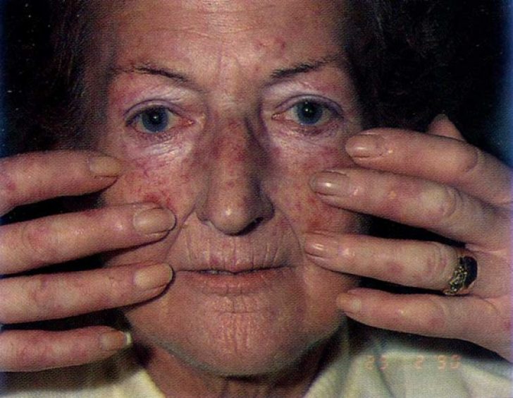
Visceral view
For him, the hallmark is the defeat of exclusively internal organs.
The jugular is discretely saturated, and its heart sounds are slightly arrhythmic. Your blood pressure is normal, and your radial pulse. The abdomen, bulky, difficult to palpate, shows a 2 cm hepatomegaly without any other visceromegaly. He has varicose veins in his lower extremities and an ulcer on the back of his right leg, not septic, with granulation tissue. The rest of the test is negative. In additional studies, it is clear that complete blood count, RBC sedimentation, glycemia, creatinine, total and fractionated proteins, prothrombin time, and liver enzymes are normal.
A separate type is considered the juvenile form of scleroderma - this is when the disease develops in childhood.
Reasons for the development of scleroderma
The true causes of the emergence and development of the considered autoimmune disease have not yet been established. Doctors have only assumptions that scleroderma has a hereditary / genetic etiology. It is interesting that for a long time the pathology may not manifest itself in any way and not even progress - it seems to “lurk” in the body. Provoke the development of pathology can be hormonal disorders, acute respiratory infectious diseases, any inflammatory / infectious process in the body that occurs in a chronic form.
Serological studies cannot be performed. The silhouette of the heart is normal. The presence of isolated atrial premature beats is observed on the ECG, and the echocardiogram shows diastolic function overload without systolic compromise. The esophagogram shows a somewhat refined esophagus, but no other changes.
The patient was treated with diuretics, vasodilators and nitrites, with improvement within one week. Clinical examination of this patient revealed a diagnosis of the etiology of right heart failure in a person with systemic disease that appeared to be consistent with his appearance, its stigmas, scleroderma type collagenase. Pathologies such as hypothyroidism, nephrosis, liver failure of any etiology were excluded, and it was believed that he suffered from collagenosis throughout the clinical condition.
In addition, injuries, hypothermia, regular poisoning of the body with chemicals, and long-term use of certain medications can serve as a "push" to the progressive course of scleroderma.
Clinical picture
The disease under consideration is quite complex - even its clinical picture is multifaceted and may vary depending on the state of the general health of the person, the level of nutritional value and other external factors. But in any case, scleroderma has common symptoms:
In the case study, other causes of pulmonary hypertension were excluded due to lack of clinical evidence and proper heart failure due to valvular damage or other causes. After examining the normal analysis of the patient, the authors performed a skin and muscle biopsy and the patient was discharged to await the result by consultation, and finally this study offered a definitive diagnosis that explains the entire clinical picture. He had an obvious symptomatic improvement with the applied treatment and a good evolution.
- spontaneous weight loss - this occurs even with excellent appetite and complete absence of physical activity;
- general weakness and fatigue - this condition is noted by patients as permanent, many doctors diagnose chronic fatigue syndrome and prescribe completely inadequate treatment;
- irregular - they are not associated with any colds and infectious diseases.
All other symptoms of scleroderma are directly related to what part of the body is affected by scleroderma.
Again, adequate clinical examination has been shown to yield a greater diagnosis; Even when the clinical picture points to a particular entity, we cannot afford to overcome obstacles to results, because, however rare, disease may occur if they are described, because they appear, of course, whenever they are suspected and looking for.
Harrison Principles of Internal Medicine. Cecil's textbook of medicine. 22nd ed. Presence of dominant T cell clones in the peripheral blood of patients with collagen vascular disorders: a prospective study of 97 cases. Clinical and serological heterogeneity in patients with antigenthromic antibodies. Pulmonary artery Hypertension associated with connective tissue diseases. Prevention of pulmonary hypertension in systemic scleroderma - a cohort study of 67 patients. Since scleroderma is called a group of diseases and syndromes that have general characteristics, it is hardening and thickening of the skin.
Skin lesions in patients with scleroderma are divided into several types:
- Dense edema- the skin acquires a very dense texture, it is impossible to fold it, if you press on the affected area of the skin, a hole will remain. Wrinkles are smoothed out on the face, but this does not bode well - soon, if ignored medical care, the face acquires an absolutely zero mimic mask. If the hands are affected, then the fingers become swollen/dense and it becomes impossible to even bend them.
- Complete or partial change in skin color- alternating areas of skin with a high level of pigmentation and its complete absence are noticeable. As the disease progresses, vascular "patterns" appear.
- Skin thinning- Patients develop deep folds around the mouth, which cause pain and make it difficult to open the mouth. In general, the skin becomes shiny, flabby and wrinkled.
In this modality of entity, it is typical that Raynaud's syndrome precedes for many years the presentation of the remaining symptoms of the disease. Manifestations of interstitial pulmonary fibrosis, clinically confirmed by bibasal crackles, without any other clinical expression, may appear, and this causes pulmonary hypertension and myocardial insufficiency. It is a rare condition, more common in women than men between the ages of 35 and 50. This is a rare entity, but when there is clinical evidence, we should insist on a positive diagnosis.
In the event that the disease in question is actively progressing and the patient does not receive any medical care, atrophied areas of the skin appear.
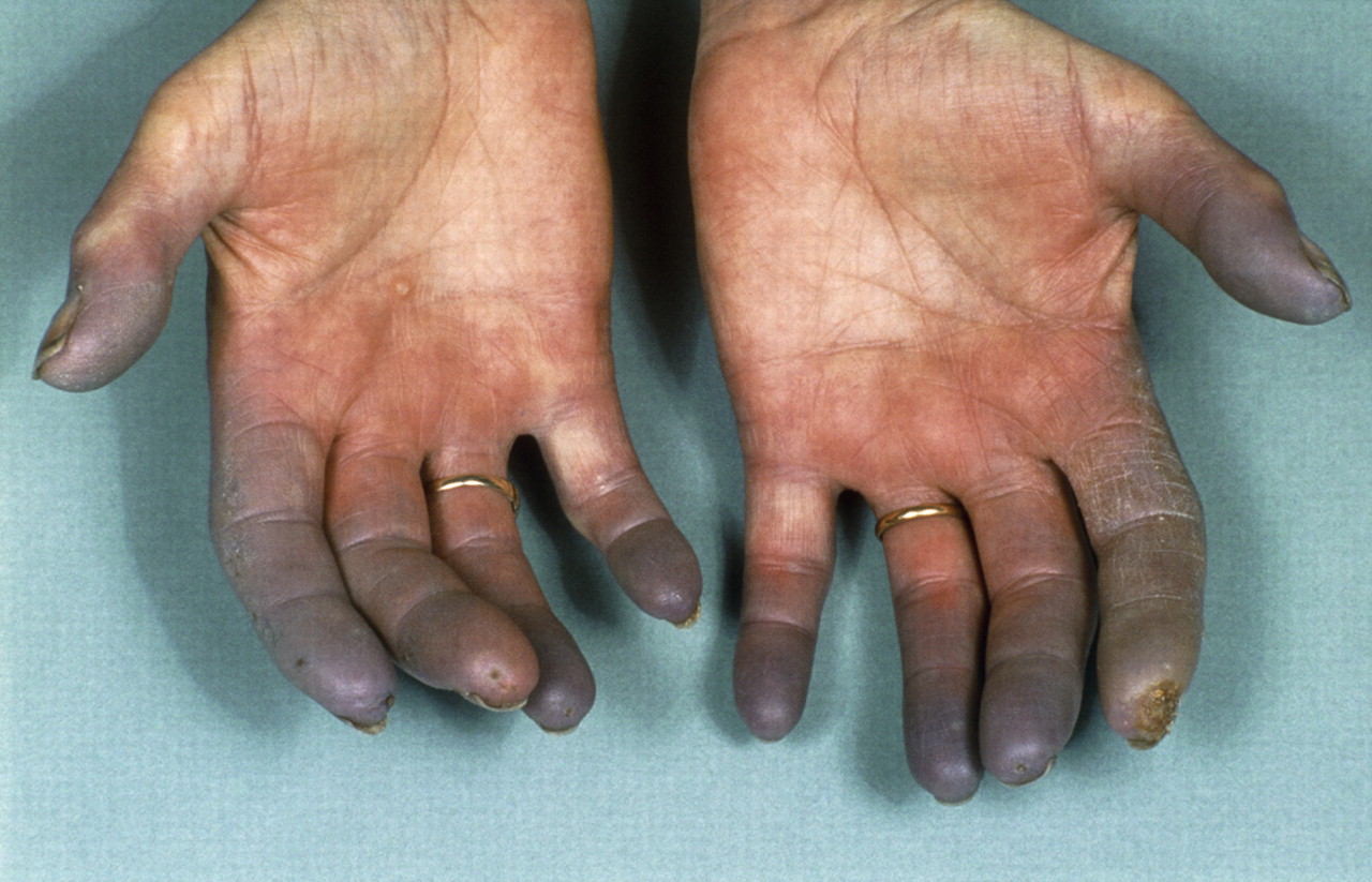
One of the pronounced signs of systemic scleroderma is Raynaud's syndrome, and often it is the only symptom. In this case, the clinical picture of the disease in question will be completely identical to the signs of Raynaud's syndrome:
This is an interesting case because she presented with pulmonary hypertension, pulmonary and developed successfully. Raynaud's disease is a rare disease that affects the arteries or blood vessels. It is sometimes called the phenomenon or Raynaud's syndrome. Arteries carry blood from the heart to other areas of your body. Raynaud's disease occurs when blood vessels narrow. This changes the amount of blood that flows into the skin, especially on the fingers and toes.
Raynaud's disease symptoms
When the vessels are narrow, they restrict or obstruct blood circulation. Lack of blood causes your skin to turn white or blue. You may also feel cold, numbness, or pain in the affected areas. Raynaud's disease is primarily associated with fingers or toes. In rare cases, it may affect the nose, ears, nipples, or lips.
- even with a slight effect of cold or any psycho-emotional “shakes”, the lumen of the vessels narrows. The result is blanching and numbness of the fingers, and after a short period of time - burning and tingling;
- if the vascular spasm persists, then the fingers begin to acquire a bluish tint, and the patient begins to feel quite intense pain;
- when carrying out any warming procedures, the pain recedes, the skin turns red.
Note:if Raynaud's syndrome is actively progressing, there are no prescriptions for drugs from doctors, then with a probability of 89%, painful and slowly healing ulcers on the fingertips will appear over time, and eventually their necrosis develops.
Raynaud's episodes can last for minutes or hours. They may be frequent or widespread. Certain events or environments may trigger them. The disease may affect one finger or foot or spread to others. When finished, your skin will turn red as the blood returns. You may feel a tingle or thud as it heats up again. Blood flow can take up to 15 minutes to return to normal.
What are the causes of Raynaud's disease?
Some people who have Raynaud's may develop skin sores or infections. This occurs when the episodes continue for long periods of time or reoccur. Few people have long-term tissue damage caused by the disease. There are two types of Raynaud's disease.
Problems in the musculoskeletal system
These include:
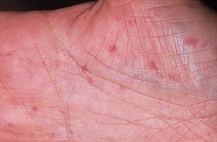
- calcification- in the area of the fingers of the upper extremities and the anatomical location of the joints, calcium salts are deposited. Outwardly, this is manifested by white foci that are clearly visible through the skin;
- muscle tissues- the patient is constantly worried about pain in the muscles, which are not associated with physical activity;
- joints large and small- the patient complains of pain in them of a aching nature, in the morning the severity of flexion / extension of the joints is noted, with the course of the disease, stiffness of movements develops;
- bones- progressive scleroderma leads to deformation of the fingers, they can be shortened and bent.
Lung injury
Symptoms of lung dysfunction may appear one of the first when other signs of scleroderma are absent. The most characteristic symptom of lung involvement will be shortness of breath and a prolonged dry cough. But in some cases, more complex pathologies develop:
Primary Raynaud's disease is more common. This is about 80% of cases. People who have this type of Raynaud's usually have mild symptoms. It can be treated with lifestyle changes. Secondary Raynaud's disease is more serious. This may require lifestyle changes and treatment. Other health problems, medications, or injuries can cause this type of Raynaud's disease. Some health problems.
Some medications can cause secondary Raynaud's disease. Some cold medicines that constrict blood vessels, such as those containing pseudoephedrine.
- Medicines containing estrogen, such as birth control pills.
- Chemotherapy drugs such as cisplatin or vinblastine.
- inflammation of the pleura- chronic pleurisy;
- inflammation of the interstitial tissue of the lungs, which provokes fibrosis - interstitial fibrosis of the lungs;
- sustained increase in the pulmonary artery- pulmonary hypertension.
Heart lesions
Patients with developing scleroderma complain of recurrent pain in the upper chest, the appearance of edema with moderate fluid intake, and increased heart rate at rest. A prolonged process of heart damage can lead to:
How is Raynaud's disease diagnosed?
Secondary Raynaud's usually occurs in people over 30 years of age. Your doctor will perform a physical examination and questions about your symptoms to help diagnose Raynaud's disease. The doctor may trigger an episode of cold water or air to see your response. You will also be asked if your family has Raynaud's disease or related health problems.
Can Raynaud's disease be prevented or avoided?
Your doctor may check the blood vessels in your nails. A blood test can help determine the type of illness or other problems that are causing the symptoms. Raynaud's disease cannot be avoided. If you have a related health problem, see your doctor to see if you have Raynaud's disease. Other factors that may affect the risk of Raynaud's disease include.
- fibrosis of the heart muscle- its compaction occurs, which provokes a violation of activity;
- endocarditis- inflammation of the inner lining of the heart;
- pericarditis- an inflammatory process occurring in the serous membrane.
Kidney damage
It can develop in two forms:
- acute form- the pathological process begins suddenly, accompanied by rapidly developing renal failure, actively rising to critical levels of blood pressure, a significant decrease in the amount of urine excreted, confusion;
- chronic form of injury- the inflammatory process in the kidneys proceeds for a long time, without any pronounced symptoms. The study of biomaterials reveals a large amount of protein in the urine, an increase in the number of red blood cells.
Problems in the gastrointestinal tract
With progressive scleroderma, lesions of the esophagus are most often detected. They are manifested by belching, heartburn, reflux of the contents of the stomach into the esophagus, difficulty in passing food into the stomach, and the formation of trophic ulcers on the mucous surface of the esophagus.
Raynaud's disease treatment
Certain medications for hands or feet in colder climates than exposure to chemicals associated with repetitive motion or vibration. This can damage certain nerves that control blood flow in your body. Family history. . Talk to your doctor about treatment options. They vary depending on the type of Raynaud's disease and general condition health.
You can make lifestyle changes to reduce episodes and symptoms. Reduce exposure to extreme cold or extreme temperature changes. Types of medications that may help. These medicines relax small blood vessels so they can open up again.
- Some calcium channel blockers.
- They also help reduce the frequency and duration of episodes.
- Alpha blockers.
As a rule, against the background of scleroderma, pathological changes in the functionality and structure of the stomach and duodenum are not detected. But there will be some violations of the intestines - weakened peristalsis, a constant feeling of heaviness in the stomach, constipation.
In addition to the above signs of the development of scleroderma, it is possible to note the emerging disturbances in the functioning of the thyroid gland (its functionality decreases) and a pronounced decrease in sensitivity in the fingers and some areas of the skin - this is how a violation in the functioning of the nervous system manifests itself.
To establish an accurate diagnosis, the patient's complaints alone will not be enough for the doctor - many of the clinical manifestations of scleroderma are identical in relation to the signs of other pathologies. Diagnostic measures are as follows:
- questioning the patient to clarify complaints - how long ago they appeared, with what intensity they proceed, whether there are violations in the work of internal organs;
- examination of the patient - with actively progressive scleroderma, the doctor may pay attention to white lesions around the joints and on the fingers, thinning and discoloration of the skin, swelling;
- - ESR will be increased, the number of leukocytes will be increased, which indicates the current inflammatory process;
- immunological studies - autoantibodies will be detected in the blood;
- histological examination of areas of the affected skin (biopsy) - this allows you to identify the current inflammatory process at the site of examination;
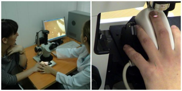
- examination of the vessels of the nail bed, capillaroscopy - the doctor can establish violations in the vascular system.
If the question of scleroderma is no longer raised, then the doctor necessarily sends the patient for an x-ray examination - this allows you to identify deposits of calcium salts that destroy processes in the nail phalanges, inflammatory processes in the lungs.
In case of patient complaints about violations in the work of the heart, an electrocardiogram, an ultrasound examination is prescribed.
Treatment methods for scleroderma
Scleroderma is treated only with complex therapy - it is not recommended to exclude something from it. It is important to seek medical attention in a timely manner in order to receive adequate treatment.
Medical treatment
The doctor prescribes medications only on an individual basis - a lot depends on the stage of development of scleroderma, what lesions are present at the time of the appointment of therapy. The main types of drug therapy are listed below.
Anti-inflammatory therapy
An excellent effect in the treatment of scleroderma is given by cytostatics, non-steroidal anti-inflammatory drugs, aminoquinoline drugs, glucocorticosteroid hormones. Proper use of these medicines can reduce the level of development of the inflammatory process, stop its progression, and reduce the intensity of the pain syndrome.
Note:many of the drugs listed above can provoke the development of severe side effects: and, manifestations of intoxication.
Therapy of disorders in the vascular system
The patient must be prescribed vasodilating drugs that improve blood circulation and reduce the level of its coagulability.
Therapy aimed at curbing the development of connective tissue compaction processes
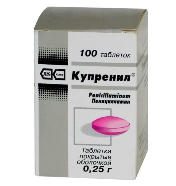 Such treatment is advisable in the later stages of the development of scleroderma, when severe lesions of the musculoskeletal system and muscle tissues are observed. Specialists most often use Kuprenil - this drug can suppress the formation of excess connective tissue and compaction.
In addition, patients take enzyme preparations, attend plasmapheresis procedures.
Note:signs of scleroderma are successfully removed with radon and / or sulfide baths - only the attending physician can make specific appointments, since this depends on the stage of development of scleroderma. AT recent times light treatment is widely used - for this, ultraviolet A is used.
Traditional medicine in the treatment of scleroderma
Important: treatment of scleroderma with folk remedies without the use of medications is impossible - in any case, the patient must visit a doctor to monitor his health, take courses of drug therapy. But official medicine does not deny the beneficial effects of folk remedies, at least they will help strengthen the immune system and increase the body's resistance.
There are many folk remedies that healers recommend taking with diagnosed scleroderma - they all deserve attention from the patient. Below are just a few of the most accessible:

Pay attention to some clarifications:
- in case of violations in the work of the kidneys, in the prepared decoction of adonis and cyanosis, you need to add a teaspoon of lingonberry leaf and the same amount of bearberry;
- with pulmonary pathology - a teaspoon of wild rosemary;
- with the appearance of blood in the urine - a teaspoon of stinging nettle and yarrow;
- with dysfunction of the gastrointestinal tract - a teaspoon of wormwood and three-leaf watch.
- when adding additional components, it is necessary to increase the amount of liquid by 300 ml.
During periods of subsidence of scleroderma, when the clinical picture becomes less pronounced, you can take herbal decoctions to strengthen immunity and reduce the level inflammatory processes. To do this, you need to prepare two different herbal collections:
- leaves of meadow geranium, lingonberry, golden rod, highlander bird, chamomile, perforated hypericum, meadow clover, plantain, dandelion roots;
- dandelion and burdock roots, calendula and medicinal chamomile flowers, grass, St. John's wort, boletus, wormwood, raspberry and plantain leaves.
Scheme for the preparation of medicinal decoctions: take two tablespoons of the desired herbal collection, pour 1 liter of boiling water and infuse the product in a thermos for at least 8 hours.
 Scheme of use: ¼ cup of decoction three times a day 30 minutes before meals. The course of treatment is 3 months, then a break of 14 days is taken and the course of using the second composition of the herbal collection also begins for 90 days.
To reduce the intensity of skin manifestations of scleroderma, you need to prepare a tincture of one part of licorice root, birch buds, cinnamon bark, wormwood and three parts of green walnuts (specific proportions: 50 g of these plant parts and 150 g of nuts). All this is poured with alcohol in the amount of 500 ml and infused for a month. Next, you should lubricate the affected areas of the skin with tincture 2-3 times a day for 3 months.
Note:treatment of scleroderma is a long process that should not be interrupted in any case. Only perseverance and regularity in the use of all the above remedies can give a sustainable result, namely, a long-term remission.
Nutrition for scleroderma
The considered systemic disease requires obligatory adherence to dietary nutrition. The task of correcting the diet is to ensure a constant supply of a sufficient amount of vitamins and minerals to the patient's body. At the same time, nutrition should be balanced and in no case "one-sided". When prescribing a diet for a patient with scleroderma, a nutritionist will definitely recommend:
- Use- it supports the immune system and normalizes the condition of the skin. Foods that are high in vitamin A include carrots, broccoli, and tomatoes.
- Ensure entry into the body- it reduces the degree of development of inflammatory processes, prevents the development of infections. Foods high in vitamin E: nut butter, all types of vegetable oils, spinach,.
- Try to eat foods that are rich in- citrus fruits, strawberries, broccoli. This vitamin protects the skin from the progression of damage inherent in scleroderma.
Nutrition for scleroderma should be varied, it is better to cook dishes with vegetable oils, consume a large amount of carbohydrates, which are found in fruits and vegetables, grains. But at the same time, obesity should not be allowed - it is already difficult for the body to deal with pathological lesions.
This is a disease characterized by damage to the connective tissue of the skin and internal organs (esophagus, lungs, musculoskeletal system, gastrointestinal tract, heart, kidneys). Scleroderma is characterized by a predominance of fibro-sclerotic and vascular changes. The disease can be localized and systemic. The first type affects only the skin, while the heart, lungs, kidneys and organs of the gastrointestinal tract affect the second type of disease with the medical term scleroderma.
Symptoms of the development of scleroderma
This disease is a progressive connective tissue disease with changes in the skin, musculoskeletal system, internal organs and widespread vasospastic disorders, vascular lesions of the type of obliterating endarteritis.
Histologically, with symptoms of scleroderma in the early stages of the process, edema of collagen fibers, an inflammatory reaction with a perivascular or diffuse infiltrate, consisting mainly of lymphocytes with an admixture of plasmocytes, histiocytes and a small number of eosinophils, are observed in the dermis. In the stage of sclerosis, inflammation disappears, and bundles of collagen fibers become homogenized and hyalinized.
The diagnosis is made on the basis of the clinical picture. From laboratory methods for limited scleroderma highest value has a histological examination, with diffuse, in addition, the detection of antinuclear and anticentromeric antibodies, the nucleolar type of luminescence in the immunofluorescence reaction. The prognosis depends on the stage and form of the disease. It is the least favorable in the systemic form of the disease, especially in the generalized form, accompanied by damage to many internal organs, which often leads to death. With limited scleroderma, the prognosis is good in most cases.
The main syndromes of scleroderma: allergic, inflammatory, metabolic disorders of the connective tissue, microcirculation disorders.
Forms of scleroderma and its manifestations
Distinguish
limited (plaque, linear),
atrophoderma Pasini-Pierini,
white spot disease – lichen sclerosus et atrophicans, and
systemic scleroderma.
plaque form of the disease (sclerodermia en plaques, morphea). The most common form of limited scleroderma, characterized by the presence of single or multiple foci of various sizes (1–15 cm or more), oval, round or irregular outlines, located on the trunk and limbs, sometimes unilaterally. In its development, the focus goes through 3 stages: erythema, induration and atrophy. The stage of erythema is hardly noticeable to the patient, since there are no subjective sensations, erythema is slightly inflammatory, bluish-pink in color.
Then, in the central zone, with symptoms of scleroderma of this form, a surface seal appears, which acquires a waxy white color (like ivory), along the periphery of which a narrow lilac rim is visible, the presence of which indicates the continued activity of the process. On the surface of individual foci there may be blisters, sometimes with hemorrhagic contents. The appearance of bubbles is associated with a violation of carbohydrate metabolism. When the focus regresses, atrophy and hyperpigmentation remain.
Linear form (sclerodermia linearis) occurs less frequently. It usually occurs in childhood, mainly in girls. Foci of scleroderma of this form can be located on the limbs (sclerodermia striata), causing atrophy of deep tissues, including muscles and bones, limiting movement if the sclerosis band captures the joints; on the penis (sclerodermia annularis) in the form of a ring in the head groove; on the scalp, often with a transition to the skin of the forehead, nose, accompanied by severe atrophy not only of the skin, but also of the underlying tissues, which makes them look like a scar after a saber strike (sclerodermia en coup de sabre).
White spot disease (lichen sclerosus et atrophicans)- a teardrop-shaped variant of limited scleroderma, but this is not generally recognized. It is characterized by small atrophic lesions of a whitish color with thin folded atrophied skin surrounded by a narrow erythematous corolla. Small foci are grouped, forming lesions up to 10 cm in diameter or more.
Atrophoderma idiopathic Pasini-Pierini manifested by several lesions, located mainly on the trunk, without or with slight induration, pinkish-cyanotic color, then replaced by a brownish tint and barely noticeable superficial atrophy. At the same time, various forms of limited scleroderma can exist.
Signs of systemic scleroderma
Systemic (diffuse) scleroderma manifested by the defeat of the entire skin (diffuse scleroderma), which becomes edematous, dense, inactive, waxy, or in the form of acrosclerosis with the most significant changes in the skin of the face and distal extremities, mainly the upper ones. The process also has 3 stages - edema, sclerosis and atrophy. Edema begins and is more pronounced on the trunk, from where it then spreads to other parts of the body.
Gradually, with the symptoms of scleroderma of this form, a seal develops. The face becomes amimic, resembles a mask, purse-like folds form around the mouth. The tongue protrudes with difficulty due to sclerosis of the frenulum. Difficulty swallowing food (narrowing of the esophagus). This kind of lesion can be localized on the genitals, in large skin folds, on the trunk. Thickened skin over the joints makes it difficult to move the fingers (sclerodactia), and is easily injured, which can lead to ulcers that are difficult to heal.
The stage of edema and compaction is replaced by atrophy of the skin and muscles. The consequence of atrophic skin changes may be poikiloderma (with telangiectasias, intermittent areas of hyper- and depigmentation and atrophy), hair loss. In a significant proportion of patients (up to 25%), there is a deposition of calcium salts in the skin and subcutaneous tissue (Thibierge-Weissenbach syndrome), Raynaud's phenomenon. From the internal organs with symptoms of scleroderma of this form, the digestive tract, especially the esophagus, as well as the lungs, heart and kidneys are mainly affected.
How to treat scleroderma with traditional methods?
Treatment should be comprehensive and aimed at suppressing the activity of immune and autoimmune reactions, intensive collagen formation, as well as normalizing the function of some of the most affected organs and systems. Medical therapy includes corticosteroid, immunosuppressive, non-steroidal, anti-inflammatory drugs, as well as vasodilators and other drugs.
How to treat systemic scleroderma?
At systemic scleroderma at an early stage, Penicillin is prescribed at 1–1.5 million units per day for 24 days, Lidase at 64 units intramuscularly every other day, for a course of 12–15 injections (4–6 courses), antihistamines, antiserotonin drugs (Diazolin, Peritol), as well as improving microcirculation and tissue metabolism (Teonicol, Reserpine, Pentoxifylline, Cinnarizine) for 2-3 weeks.
After the main course of treatment, Prodectin or Parmidin is prescribed for a month, Andecalin 10–40 units intramuscularly (for 2–4 weeks), vitamins, especially A and E, biogenic preparations (aloe, vitreous body, ATP, etc.), Solcoseryl , Actovegin. With a pronounced activity of the process and significant immune disorders, it is necessary to treat scleroderma using hyperbaric oxygenation, plasmapheresis, hemosorption, and corticosteroids. Usually, in small doses (Prednisolone 20-40 mg every other day with a gradual decrease in dose after reaching a clinical effect to maintenance).
Non-steroidal anti-inflammatory drugs, cytostatics are also used (for example, Azathioprine or Cyclophosphamide but 100-150 mg per day, Methyldopa 0.5-2.0 g per day).
One of the basic therapy drugs is Kuprenil. Treatment in the hospital begins with a small dose of 0.15-0.3 g per day, which is increased weekly by 0.15 g to a daily dose of 1-2 g. The drug at this dose is used for several months (on average, about 6), then it is reduced by 0.15 g per week to maintenance - 0.3–0.6 g, which is continued for a long time, at least a year.
With Raynaud's syndrome, calcium antagonists (for example, nifedipine) are indicated, with calcification - disodium salt of ethylenediamine tetraacetic acid (EDTA). Useful physiotherapy (warm baths, paraffin, mud), gymnastics, massage.
How to treat localized scleroderma?
At limited scleroderma repeated courses of Penicillin are prescribed in combination with Lidaza 64 IU / m daily or every other day up to 20 injections, vasoactive drugs. In some patients, Lelagil is effective (0.25 g once a day), small doses of Kuprenil (0.45 g per day), prescribed for several months. It is recommended to lubricate the foci with corticosteroid ointments (Hydrocortisone, Prednisolone), Solcoseryl, Indovazin, Heparin, Indomethacin ointment, Troxevasin gel.
The usefulness in the treatment of scleroderma of this form of Carnitine chloride (5 ml of a 20% solution) 2 times a day orally for 35–45 days, with repeated courses after 1–4 months, was noted; in the interval between courses, it is appropriate to prescribe Dipromonium 0.02 g 3 times a day, 30-40 days): Diucifon (0.1-2 g per day orally in 5-day cycles with one-day breaks, 4-6 cycles per course); Unitiol (5 ml of a 5% solution once a day, 5–20 injections per course; Taktivin or Timoptin (at the rate of 5–10 mcg per 1 kg of body weight, subcutaneously on days 1.5, 10, 15 and 21, for a course of 500–550 mcg); Tigazon (at the rate of 1 mg per 1 kg of body weight for 2–3 weeks, then 0.6–0.8 mg per 1 kg of body weight for 4 weeks with a gradual decrease in dose to 25 mg per day and drug withdrawal approximately 2 weeks).
Treatment of linear scleroderma
At linear scleroderma Phenytoin is prescribed (initially, 0.1 g 2-3 times a day, then for a long time, 0.1 g per day), antimalarial drugs (for example, Delagil, 0.25 g per day). There is evidence of the positive effect of radon baths, Dimexide (in pure form or in a 30–90% solution, including with corticosteroids, such as Dexamethasone at 0.05% concentration).
On the foci in the treatment of scleroderma, it is advisable to use a 20% solution of Lidase or Ronidase, proteolytic enzymes using phonophoresis. It is possible to use Bernard's diadynamic currents, local baro- and vacuum therapy, trypsin and chymotrypsin (in the form of intramuscular injections or administered using ultrasound), laser beams (helium-neon or infrared), microwave electromagnetic field in alternation with iodine-bromine baths, electric and phonophoresis of Ronidase, Lidase, Potassium Iodide, Ichthyol; applications of Paraffin, Ozokerite, therapeutic mud, Naftalan.
Dispensary observation is important to maintain the therapeutic effect; repeated sanatorium treatment at balneological and mud resorts, massage, physiotherapy; from a year 2-3 courses of Lidaza, biogenic preparations, alternating them with drugs that improve microcirculation, vitamins, in combination (if necessary) with external agents (Dimexide, Hydrocortisone ointment, electrophoresis with Lidaza).
Physiotherapy for scleroderma
Physical methods are aimed at reducing the immune response (immunosuppressive methods), stopping inflammation (anti-inflammatory methods), restoring connective tissue metabolism (fibromodulating methods) and microcirculation disorders (vasodilating methods). These tasks help to implement the following methods of physiotherapy:
Immunosuppressive methods: aerocryotherapy, drug electrophoresis of immunosuppressants, nitrogen baths.
Anti-inflammatory methods of treatment: UHF-therapy on the area of the adrenal glands, hydrocortisone ultraphonophoresis.
Fibromodulatory methods: pelotherapy, hydrogen sulfide, radon baths.
Vasodilating methods: paraffin therapy, ozokeritotherapy.
Contraindications to treatment: acute course of the disease with a high degree of activity, severe damage to the heart, kidneys, peripheral and central nervous system.
Sanatorium-resort method of treatment of scleroderma
Patients with systemic scleroderma with subacute and chronic course with minimal activity of the process are sent to balneological resorts with hydrogen sulfide waters (Yeisk, Sergievsky Mineralnye Vody, Pyatigorsk, Sochi, Belokurikha, Truskavets, Bakirovo, Goryachiy Klyuch, Novye Klyuchi, Ust-Kachka, Klenovaya Gora, Khilovo , Argman, Surakhany, Chimion, Baldone, Shikhovo).
Contraindications to the spa treatment of scleroderma are:
acute course of the process,
high degree of activity
severe damage to internal organs.
Physioprophylaxis is aimed at suppressing intense collagen formation (fibromodulating methods), the activity of immune and autoimmune reactions (immunosuppressive methods), as well as mobilization defensive forces organism and hardening (catabolic methods).
Folk remedies and recipes for scleroderma
Before treating scleroderma, it is necessary to be diagnosed in a hospital, as improper treatment can lead to complications. Drug treatment can be supplemented with folk remedies, but remember that only supplement, not replace.
You can use a compress. To do this, bake a small onion in the oven, then chop and add a teaspoon of honey and two tablespoons of kefir. Mix thoroughly and compress overnight four times a week.
One way is herbal decoction. It is necessary to mix lungwort, knotweed and field horsetail in equal proportions. Grind everything and pour one tablespoon with a glass of water and put in a water bath for 15 minutes. Infuse for half an hour, after which you can take. Schedule - three times a day for the third part of the glass half an hour before meals or an hour after.
If there are complications during scleroderma, the following will help folk remedy. Gather the leaves of peppermint, lingonberry, plantain, raspberry, medicinal sweet clover, meadow geranium, St.
Some of these ingredients can be bought at a pharmacy, some can be assembled independently. Mix all the herbs in equal amounts, grind, then pour two tablespoons of herbal powder with a liter of boiling water and leave to infuse overnight in a thermos. In the morning, strain the infusion and drink three times a day a quarter cup half an hour before meals. The course lasts three months.
Herbal medicine can offer you many recipes, but before using them, be sure to consult your doctor.
Causes of scleroderma
The causes of scleroderma are usually hypothermia, all kinds of infections of the nervous system, industrial vibration. All this contributes to a change in the walls of blood vessels, as a result of which they become denser and lose elasticity, which may cause the lumen of small vessels to close. All these changes disrupt the blood supply to tissues and organs. Scleroderma may also have a genetic predisposition.
Predisposing and provoking factors of scleroderma include:
hypothermia,
acute or chronic infections,
sensitization,
endocrine dysfunctions (hypoestrogenia, hypocorticism).
The main mechanism for the development of scleroderma lies in the violation of collagen synthesis and metabolism, which is confirmed by increased activity of fibroblasts in tissue culture, increased collagen production in the active phase of the disease, and high excretion of hydroxyproline. Intensive production of immature collagen by fibroblasts leads to disruption in the microvasculature. This is facilitated by neuromuscular dysfunction, defects in the immune system, confirmed by the presence of autoantibodies (antinuclear, anticentromeric to RNA, DNA, etc.), immune complexes, emerging immunodeficiency with signs of cell-mediated hypersensitivity.
In the development of scleroderma, the participation of histamine and serotonin in the formation of edema and microcirculatory disorders, the effect of an increased content of acid mucopolysaccharides in the dermis on connective tissue sclerosis, the role of hereditary factors, as evidenced by family cases, the association of the disease with antigens of the HLA system (AL, B8, B18, B27, Bw40, DRI, DR5).